Lesson 3
Common Hoof
Disorders
Sole
Bruises
Definition
– Sole bruises are actual bruising (bleeding) under the sole surface of the
horse’s foot. (Remember – the sole is not intended to be a weight bearing part
of the bottom of the foot, it is normally concave and shouldn’t hit the ground
surface with full force.)
Symptoms – There is some sensitivity
over the bruised area especially if hoof testers or some other pressure is
applied over the bruised area. It doesn’t always cause a noticeable
lameness. Paring away of the sole in the
bruised area will show discolored (bluish-black) sole .
Causes – Concussion to the sole by hard
objects such as large rocks, gravel or hard road surfaces. Unleveled or poorly fitting horseshoes can also cause sole
bruises.
Location – the under part of the sole
surface of the hoof
Prevention – Shoeing the horse will
help protect the sole. Keep the sole of the foot in a healthy condition. Pads are used when the sole is flat or not
very tough.
Treatment – Treatment would consist of
Epsom salt soaks, toughing up the sole with hoof conditioners (iodine or koppertox) and the use of pads for protection of the
bruised area, if the horse is to continue to be used.
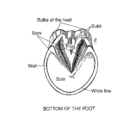
Corns
Corns are a sole bruising which occurs
in the “seat of corns,” the area at the heel formed by the angle of the bars of
the heel. See foot chart. The bruising
is usually due to a shoe that has been left on too long. The foot grows back
and the end of the shoe will put pressure on the sole area at the angle of the
bar. When the horse walks or moves on the shoe in this position, it puts
pressure in an area that is not designed to bear weight so it will easily
bruise.
Hoof
Abscess
Definition – A hoof abscess in an
infection or pus pocket on the underside of the sole or somewhere within the
sensitive part of the hoof.
Symptoms – A severe lameness, the horse
is normally reluctant to put any weight on the affected foot until the abscess
breaks and drains. There is heat in the affected foot, especially where the
abscess is located, and hoof testers or thumb pressure over the abscess will
illicit a pain response in the horse. Oftentimes there will be swelling in the
pastern area, and a strong digital pulse can easily be felt in the affected
foot.
Causes – A sole bruising, if severe
enough, can develop into a hoof (sole) abscess. The most common cause of the
hoof abscess is a puncture wound of the sole or frog that penetrates into the
sensitive tissue. White line disease or other separations of the hoof area that
allow bacteria to penetrate into the sensitive structures of the hoof can cause
a hoof abscess. “Quicking”
a horse (driving a nail into the quick, or sensitive
part of the foot) can cause an abscess.
Location – An abscess can occur
anywhere below the hoof capsule in the sensitive part of the foot, but usually
follows the path of least resistance, so many will travel to the white line,
then break out at the coronary band.
Prevention – Keeping the foot healthy,
clean and dry, will help prevent hoof abscesses from developing.
If the horse is “quicked,” or gets a puncture
wound, paring it out with a hoof knife, or treating it immediately with strong
iodine may help
prevent a hoof abscess from developing. Putting the horse on
preventative antibiotics may also be warranted. Seek advice from your
veterinarian anytime you see blood coming from a hoof puncture.
Treatment - Hot Epsom salt soaks will help draw
the abscess out of the hoof, as will a poultice application under a hoof
bandage. Both procedures should be
used. Consult your veterinarian if the
abscess does not resolve within a few days.
Antibiotics may be necessary. Once the abscess breaks and drains, you
should still soak and pack for a few days, treating the open area with
something like strong iodine to toughen the area and close the opening.
Sand (hoof) Cracks (toe, quarter or heel
cracks)
Definition – a vertical crack in the
hoof wall. Cracks can be superficial (not penetrate the sensitive lamina) or
deep (penetrate the sensitive lamina and cause blood to appear at the surface.)
Symptoms – If the crack is
superficial, there are no lameness symptoms, but if the crack penetrates into
the sensitive tissue of the hoof, blood and pain will be noticed.
Cause – Dry, brittle hooves are more
prone to hoof cracks. Cracks can also occur from imbalanced feet or uneven
weight bearing. A deep wire cut or laceration into the conorary
band will produce a defect in the coronary band where the hoof grows from, and
a crack will always grow down from there for the rest of the horse’s life.
Location – the location of the hoof
crack is usually designated by toe, quarter or heel crack. They can start from
the ground surface and travel up, or they can start from the coronary band and
travel downward.
Prevention – Good hoof management
practices will help prevent hoof cracks. Keeping the hooves
healthy and pliable. Proper balancing and shoeing will also prevent hoof
cracks.
Treatment – The important aspect of
treating hoof cracks, is to stop the crack from
spreading, stabilize it the best you can, and promote healthy hoof growth to
allow the crack to grow out as quickly as possible. The crack can be grooved
with a horizontal groove in the hoof wall at the end of the crack. This might
keep it from spreading, especially if it is not a deep crack. Usually a horseshoer and /or a Veterinarian will be involved in the
treating of a hoof crack. They will use shoeing (shoes with clips to stabilize
the crack, or a shoeing technique to take direct pressure off the crack) . OR they may use staples, or other hardware (screws and
plates) or even special acrylic bonding material to repair the crack. (Remember
a horses hooves grow down about 1/4 to 1/3 of an inch
a month)
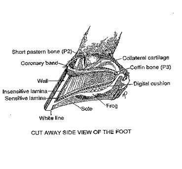
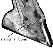
Navicular disease or syndrome
Definition – Navicular
disease is a chronic (long standing) disease involving inflammation of the navicular bone and navicular area
of the front limbs.
Symptoms – Navicular
disease usually shows as a mild to medium lameness condition of the front
limbs, usually one front being worse than the other. The horse is normally
reluctant to place his heels to the ground at a trot, and will stumble and
short stride. A head bob is usually noticeable at the trot (head goes up when
the sorest foot hits the ground) especially when going in a circle. When standing, a horse with navicular
pain may point (place one front foot slightly ahead of the other, therefore
relieving the pressure on the navicular bone).
X-rays of the navicular bone will show spurring of
the bone and /or holes in the navicular bone
(lollypop looking holes). Although occasionally a horse with clinical navicular disease will have clean x-rays. In this case the
explanation is usually that the pain is associated with the soft tissue
structures of the navicular area. (This is still
considered to be navicular disease)
Location – Navicular
bone, navicular bursa, and deep digital flexor
area over the navicular bone. Seen
almost exclusively in the front feet. Usually both front feet are
involved, one worse than the other.
Cause
- Usually seen in older horses as a wear and tear type of damage, seen in
horses with small hooves, short, straight pasterns, or low heels and long toes.
Concussion over this navicular area below the heel is
the main cause. Heredity may play a role.
Prevention – Short, straight pasterns
and small feet can predispose a horse to navicular
problems because of the increased concussion to the heel area and navicular area of the foot over time. Purchasing horses
with adequate size of foot in rel;ation
to their size, as well as those with good pastern conformation, and normal and
equal hoof to pastern axis, as well as keeping these horses shod in the correct
way, can prevent you from having problems with navicular
disease in your horses.
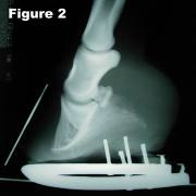
Treatment – Corrective Shoeing or
trimming – High heels, short and rolled toes . Bar shoe, bar across the
middle third of the frog. Medical management – Bute - pain management. Isoxuprine
tablets in the feed will help increase blood flow to the navicular
area.
Laminitis – Founder
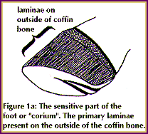
Definition – Inflammation of the
lamina of the feet.
Causes:
1.
Overeating of grain
2.
Excessive cold water ingestion when horse is not cooled out (to hot)
3.
Overeating of lush green pasture (alfalfa or other legumes)
4.
Overwhelming infection of disease.
5.
Road founder – overwork on hard ground.
6.
Overexposure to cortisone type drugs
7.
All above basically encompass over stressing a horse due to poor training or
management decisions. (Pushing a horse over what his system can tolerate) Horse
must become slowly accustomed to changes in things like feed, or exercise)
Signs: Three Phases
1.
Developmental Phase – from exposure to the cause until first symptoms appear.
If treated during this phase you may be able to prevent the laminitis.(Nsaids, Lower blood pressure,
and laxatives (mineral oil)
2.
Acute Phase – Heat, and extreme pain in affected feet (usually both front
feet). Walking on eggshells, bounding digital pulse.
3.
Chronic Phase - >48 hrs. 0r when P3 begins to rotate.
Treatment:
Acute
1.
Treat for pain and inflammation -( Bute
or Banamine)
2.
Anti-endotoxin – (Banamine)
3.
Lower systemic blood pressure (acepromazine)
4.
Anticlotting treatment (Bute
or Banamine)
5.
Treat feet: a. Lily pads or gauze rolls taped to frog area or sand or mud (frog
and sole pressure desirable)
6.
Ice first 48 hours.
Chronic
1.
Control pain.
2.
Antibiotics may be in order
3.
Diet rich in ingredients responsible for healthy hoof growth (Biotin, Zinc, and
Methionine)
4.
Corrective shoeing – (heart bar shoe, hoof resection, etc.)
Prognosis
–Guarded